Dental treatment under a microscope – what it offers for caries and when it is worth choosing
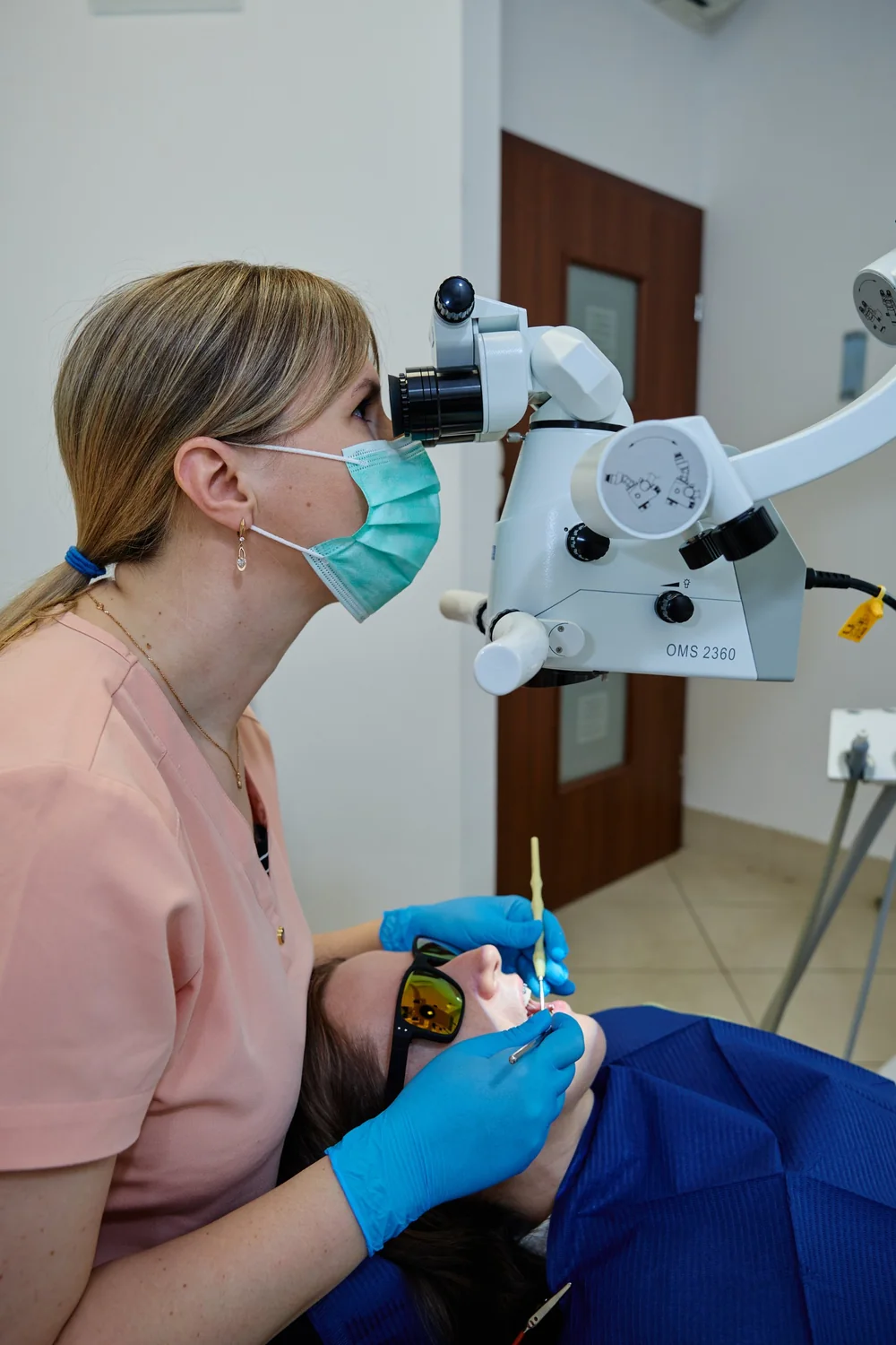
Dental treatment under a microscope is ordinary treatment, such as removing caries and placing a filling, in which the dentist works at high magnification with strong light directed deep into the cavity. The microscope helps with precision, but it is not needed for every tooth, and studies have not yet confirmed that fillings placed under magnification last longer.
This article covers caries treatment and fillings: what can be seen under a microscope, what the research says and when this option is worth considering. We describe the microscope in endodontics separately in the article on root canal treatment under a microscope (PL).
How dental treatment under a microscope works
The stages are the same as without a microscope: the dentist examines the tooth, removes the caries and restores the cavity. What changes is how clearly the working area can be seen. The operating microscope stands next to the chair on a movable arm and does not touch the patient.
Its main features are:
- adjustable magnification – studies of restorative treatment have used magnifications from about 6 to 16 times,
- coaxial light – it travels along the dentist's line of sight, so there are fewer shadows deep in the cavity,
- magnification that changes during the work – the dentist adjusts it to suit each stage of the procedure.
The microscope's light is strong, so the patient wears protective glasses during the procedure.
What dental treatment under a microscope can make easier
In restorative treatment, magnification can be particularly helpful in these situations:
- A deep cavity close to the pulp. Current practice often uses selective caries removal: the dentist thoroughly cleans the edges of the cavity and may leave some affected dentine near the pulp, sealing it in. In a review of five randomised trials, this approach was linked to fewer pulp exposures than complete caries removal. These trials did not, however, test the effect of the microscope itself.
- Replacing an old filling. Under magnification it is easier to inspect the edges of a filling and see where the material ends and the tooth begins.
- A small cavity and conservative preparation. Better visibility can help the dentist work more accurately along the intended boundary.
- A suspected crack. The microscope allows a close look at the tooth surface, but it is usually not enough as the only method for assessing cracks.
Dental treatment under a microscope, with loupes and without magnification
| No magnification | Loupes | Microscope | |
|---|---|---|---|
| Magnification | none | a few times, 2.5–5× in the studies described | adjustable, 6.4–16× in the studies described |
| Light | dental unit lamp | dental unit lamp or LED light on the loupes | coaxial |
| Preparation precision in a phantom-head study | lowest | intermediate | highest |
| Filling results after 12 months | no significant differences | no significant differences | no significant differences |
The last two rows come from two separate studies. They show that greater precision in a training setting does not necessarily translate into a measurably better filling after one year.
When dental treatment under a microscope makes sense, and when it is not needed
This option is worth discussing with your dentist when:
- the cavity is deep and you want the most careful work possible near the pulp,
- an old, leaking filling is being replaced,
- the cavity is in a hard-to-reach place,
- a crack is suspected and the dentist wants to take a closer look.
A microscope is usually not necessary for a small, clearly visible cavity. Nor will it solve problems that require a different kind of treatment:
- the tooth hurts on its own or at night – root canal treatment may be needed,
- the cavity is very large – a ceramic inlay or a crown may be a better solution,
- caries between the teeth is not visible – looking at the tooth, even under magnification, does not replace an X-ray when the dentist considers one necessary.
What dental treatment under a microscope looks like
- Examination. The dentist assesses the tooth, orders an X-ray if needed and discusses the plan and cost with you.
- Anaesthesia and isolation. Local anaesthesia is used if needed. The tooth is kept dry, e.g. with a rubber dam.
- Setting up the microscope. You receive protective glasses, and the dentist adjusts the focus and light.
- Caries removal. The dentist prepares the cavity while trying to preserve healthy tissue.
- Filling. Composite is placed in layers, cured with light and polished.
After anaesthesia, don't eat until the numbness wears off, which usually takes 1–3 hours. The tooth may be sensitive to cold for a few days. If the pain is severe or getting worse, contact the clinic.
Dental treatment under a microscope at LumiDent in Ursynów
At LumiDent, restorative dentistry in Warsaw Ursynów is provided by Dr Julia Kartynnik and Dr Rusłan Osmanov. The clinic also uses the microscope for diagnostics, root canal treatment and professional cleaning. Your dentist will tell you after the examination whether it would be useful for your tooth.
The clinic is at ul. Alternatywy 7 lok. U5, 02-775 Warsaw, and is open Monday to Saturday, 10:00–21:00.
How much dental treatment under a microscope costs
At LumiDent, using the microscope during caries treatment is a separate item added to the cost of treatment. The total cost also depends on:
- the size of the cavity and the number of tooth surfaces being restored,
- the chosen material and type of restoration,
- anaesthesia and a rubber dam,
- any diagnostics needed, such as an X-ray.
You will find current prices in the restorative dentistry and fillings price list. The dentist gives the exact cost after examining the tooth.
What research says about dental treatment under a microscope
- Precision. In a study of 16 dentists, plastic blocks were prepared in a phantom head. Work was most accurate under a microscope (6.4×), less accurate with loupes (2.5×) and least accurate without magnification. These were simulated conditions, however, not treatment of patients.
- Fillings in back teeth. A randomised trial of 130 cavities in 85 patients compared working with the naked eye, with 3× and 5× loupes and under an 8.5× microscope. After 12 months there were no statistically significant differences. The magnification groups did score higher on some criteria, but follow-up lasted only one year.
- Treatment decisions. In a laboratory study, three dentists assessed the chewing surfaces of 300 teeth. A microscope at 16× did not improve their decisions on whether to place a filling.
- Enamel cracks. In a laboratory study, a microscope (8×) performed worse than two transillumination methods, which shine light through the tooth. The authors suggest that the microscope could be combined with transillumination.
In short, a microscope improves visibility and, in simulated conditions, precision as well. There is no strong evidence, however, that it extends the life of a filling on its own. A 2026 review of endodontics also points to a lack of randomised trial evidence for an independent effect of the microscope on non-surgical treatment outcomes.
Do you have a deep cavity or an old filling to replace? Book a visit online or call +48 732 205 427.
This article is for information only and does not replace a consultation with a dentist.
Tooth decay treatment, fillings and bonding.
Questions about restorative dentistry
Does dental treatment under a microscope hurt?
The microscope does not touch the patient, so dental treatment under a microscope is no more painful than treatment without one. For deeper caries the dentist usually suggests local anaesthesia, while shallow cavities can sometimes be treated without it. Afterwards the tooth may react to cold for a few days, which usually settles on its own.
Will a filling last longer after dental treatment under a microscope?
That cannot be promised. In a clinical trial of 130 fillings in back teeth, there were no significant differences after 12 months between working with and without magnification. How long a filling lasts after dental treatment under a microscope also depends on the cavity size, isolation, your bite and oral hygiene.
Does dental treatment under a microscope take longer?
Dental treatment under a microscope can take somewhat longer, because the dentist sets up the equipment and works in close detail. Placing a composite filling itself usually takes 30 to 60 minutes, depending on the size and position of the cavity. Your dentist can estimate the visit length after examining the tooth and the extent of treatment.
Is dental treatment under a microscope needed for every cavity?
No, dental treatment under a microscope is not necessary for every cavity. A small, clearly visible cavity can usually be treated successfully without such high magnification. A microscope tends to be more useful when a cavity is deep or hard to reach, or when the edges of an old filling need close inspection. Discuss it with your dentist after the examination.
Does dental treatment under a microscope cost more?
Yes. At LumiDent, using the microscope during caries treatment is a separate item in the price list, added to the cost of the filling itself. Dental treatment under a microscope does not change the price of anaesthesia or a rubber dam, which are also charged separately. The dentist presents the total cost before starting work, after examining the tooth.
Related articles
Ceramic inlays, onlays and overlays – why they are not "ceramic fillings" and when they are used
Inlays, onlays and overlays differ in how much of the tooth they cover. When a lab-made ceramic restoration is a better choice than a large filling.
Related serviceRestorative dentistry in Warsaw Ursynów
Restorative dentistry involves removing tooth decay and rebuilding the tooth with composite material. The aim is to preserve as much healthy tooth tissue as possible.
- Eichenberger M. et al. – Effect of Magnification on the Precision of Tooth Preparation in Dentistry. Oper Dent, 2018
- Çubukcu İ., İşcan Yapar M. – The role of visual aids in the clinical success of posterior restorations: randomized controlled clinical trial. Heliyon, 2024
- Akarslan Z., Erten H. – The Use of a Microscope for Restorative Treatment Decision-making on Occlusal Surfaces. Oper Dent, 2009
- Hausdörfer T. et al. – Three Visual–Diagnostic Methods for the Detection of Enamel Cracks: An In Vitro Study. J Clin Med, 2023
- Figundio N. et al. – Deep Carious Lesions Management with Stepwise, Selective, or Non-Selective Removal in Permanent Dentition. Healthcare, 2023
- Ucmak S. – The Dental Operating Microscope in Endodontics: A Narrative Review. Cureus, 2026